


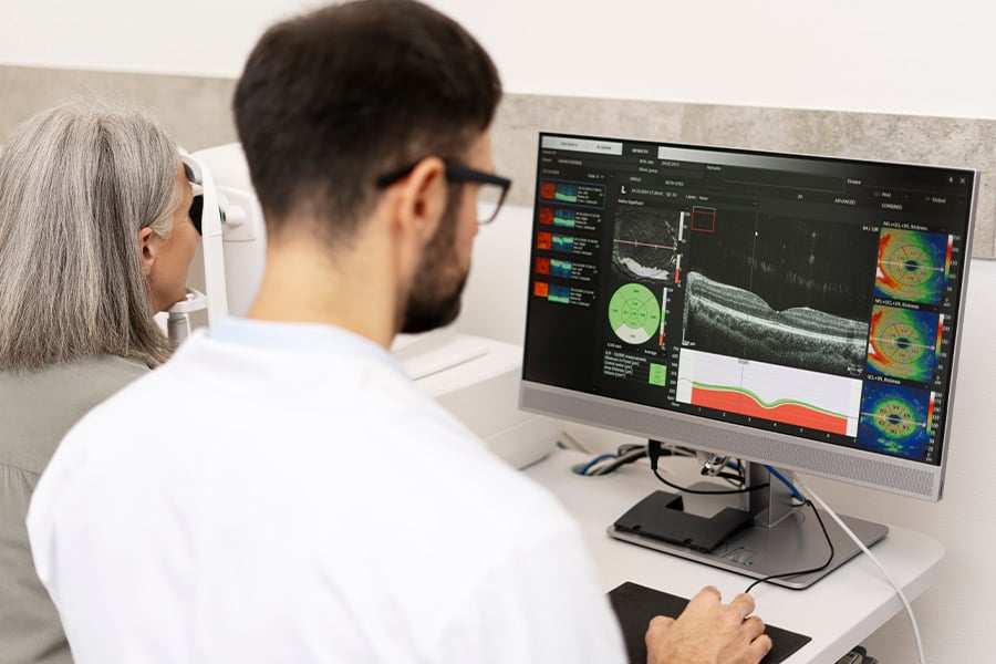
Computed tomography (CT) scans have become an essential tool in modern medicine due to their speed and ability to produce detailed images of bones, organs, and soft tissues, facilitating the timely diagnosis of diseases.
Depending on their radiation dose, CT scans are classified as standard or low-dose. Low-dose CT scans expose the patient to a lower radiation dose during the exam, although they have a significant limitation: they produce images with lower resolution and higher visual noise.
To address this challenge, the University of Santiago de Chile (Usach) conducted the R&D project “Reconstruction of Low-Radiation-Dose Computed Tomography Scans Using Accelerated Generative Models Based on Diffusion Techniques.” The project, which spanned over two years, was led by Usach Dr. Violeta Chang, a faculty member in the Department of Computer Engineering of the Faculty of Engineering and deputy director of the initiative.
Dr. Chang notes that while prior research has proposed using generative adversarial networks (GANs) and diffusion models to enhance medical image resolution, most published studies have relied on small clinical sample sizes.
“Unlike these proposals, our goal was to move toward an AI-based solution that could be validated using a clinical methodology capable of reconstructing low-radiation-dose images and bringing them closer to the quality of a conventional CT scan,” notes Dr. Chang.
Validation and Artificial Intelligence
The project brought together the capabilities of the Applied Artificial Intelligence laboratories at Usach and the Computer Vision laboratories at the University of Los Andes, forming a multidisciplinary team comprising specialists in computer science, biomedical engineering, and radiology.
Bridging academic strengths, the project combined the technical capabilities of Usach's Applied Artificial Intelligence Lab and the University of Los Andes' Computer Vision Lab. The resulting multidisciplinary collaboration brought together computer scientists, biomedical engineers, and clinical radiologists to solve complex medical imaging challenges.
Addressing information loss in low-dose CT scans requires robust testing. During the first stage, researchers simulated low-dose imaging using public CT scan databases to analyze image quality. In partnership with radiologists at Clínica Santa María, the team conducted controlled experiments to refine their findings.
Next, the researchers tested physical CT scanners and anatomical phantoms, adjusting radiation parameters to evaluate how dose levels impact visual image quality. Using these results, the team built new databases and developed a mathematical model to accurately replicate CT images at varying radiation doses.
“All this information allowed us to create artificial intelligence models designed to reconstruct low-dose images with a resolution similar to that of a conventional CT scan,” notes Dr. Violeta Chang.
Marketing Potential
Applications of this CT imaging technology promise safer scans for patients and clearer, reliable diagnostic images for radiologists and medical specialists.
Future developments could transform this technology into an online platform for medical imaging data management. “One option is for this solution to function as a cloud-based service, automatically optimizing images before medical analysis,” notes Dr. Chang.